
NPS photo.
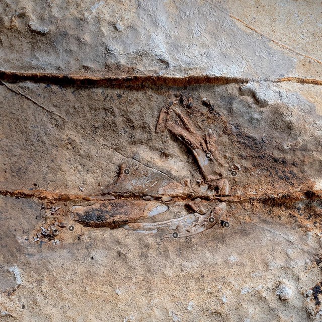
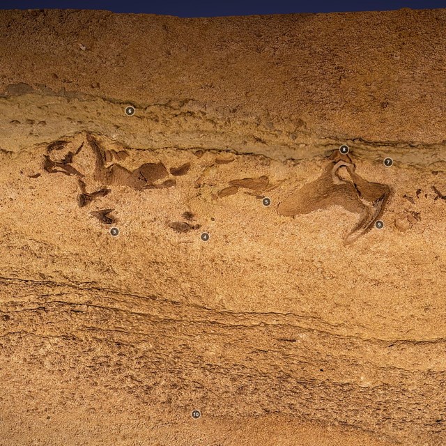
The caves of Mammoth Cave National Park have cut through the Late Mississippian Ste Genevieve Limestone, where the fossil rich Joppa member is exposed. A well know cave passage, with over 100-years of exploration was recently found to also contain the remains of a 330 million years old primative shark species. Among the fossils preserved in Mammoth Cave is a section of cartilage of the Mississippian shark Saivodus. This specimen cannot be readily collected, and casting would be very difficult. For this reason, the park decided to use photogrammetry to get a 3D reconstruction of the fossil for study and display.

NPS photo.
The discovery of shark fossils, with preserved portions of the skulls and connective tissue as well as teeth, is an exciting discovery. For such important specimens (species known until this chance discovery only from teeth) scientists would instantly begin to think about recovery and removal for analysis. However, these fossils are deep within a cave and reached through tight, sinuous and challenging passageways, that required crawling and squeezing to reach. Granted the trip started out with an elevator ride down into the cave, but that was just the first 2 minutes of the hour-long traverses to reach these sites. Travel to one of the two sites included an approx. 240 meter or 640-foot-long crawl (think more than 2 football fields) over loose and sharp gravel and gypsum crusted rock. Hence, recovery of the specimen is not an option.
Cavers and scientist often carry cameras for their explorations. For the documentation of these fossils, we also brought a computer tablet, digital controllers, lights, tripods and scale bars to then photograph these important paleo-sharks with studio-like (or nearly so) conditions. Together, all this equipment sounds like a lot, but when broken up between members of a caving team, it becomes easily transportable; unlike a single block of rock that would weigh in at 100s of pounds. We spent two days traversing cave passages with our equipment to complete the photography.

NPS photo
The original data being used for research details elements of the fossil and surrounding rock down to sand grain size, or less than 1/4-millimeter (~0.01 inches). Therefore, details in the teeth and preserved cartilage can be seen and measured. Additionally, structure within the sediments surrounding the fossil provide insights about the environment and water depth where these sharks were buried to become fossils.
Mammoth Cave Virtual Collections—Fossils in 3D
Interactive 3D Models Collected from deep within Mammoth Cave National Park.
Related Links
- Mammoth Cave Fossils
- Mammoth Cave National Park, Kentucky—[Geodiversity Atlas] [Park Home]
- NPS—National Fossil Day
- NPS—Photogrammetry
- NPS—Fossils and Paleontology
- NPS—Geology
Last updated: October 14, 2020