National Park Service Museum Program
The National Park Service manages one of the world's largest museum systems, with museum and archival collections located in in over 390 parks and centers throughout the US.
Official websites use .gov
A
.gov website belongs to an official government
organization in the United States.
Secure .gov websites use HTTPS
A
lock (
) or https:// means you've safely connected to
the .gov website. Share sensitive information only on official,
secure websites.
The National Park Service manages one of the world's largest museum systems, with museum and archival collections located in in over 390 parks and centers throughout the US.
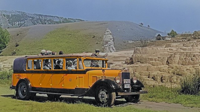
This exhibit explores travel through Yellowstone NP, from the earliest Native American groups and European Americans to present-day visitors
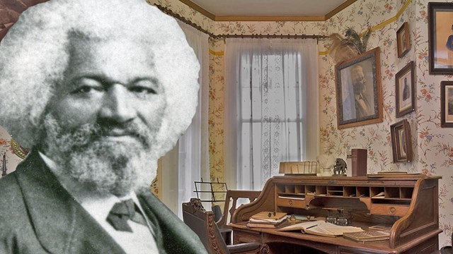
Explore thematic virtual exhibits featuring National Park Service museum collections.

Explore online museum features created by parks.
Guidance and standards on managing, preserving, protecting, documenting, and accessing museum collections.

Short, focused technical leaflets that provide practical and easy-to-use guidance on the care of museum collections.

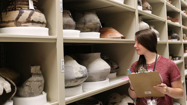
The Web Catalog is a searchable online database that provides access to thousands of images and records from the NPS museum collections.
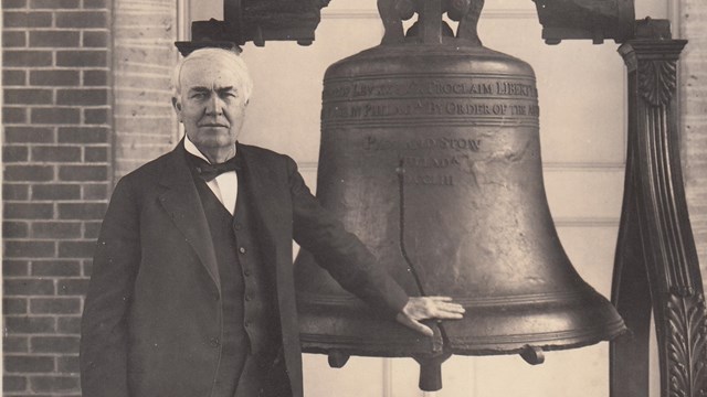
National Park Service archival collections preserve historic records and make them available for research and use.

Lesson plans use National Park Service objects in student-centered educational activities.
Last updated: December 10, 2024