
|
Geological Survey Professional Paper 58
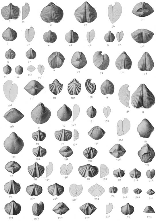
The Guadalupian Fauna |
PLATES — PLATE XIII.
CAPITAN FORMATION, GUADALUPE MOUNTAINS.
|
SPIRIFER MEXICANUS Shumard (p. 360). | |
| FIGS. 1 and 1a. |
A ventral valve of medium size. |
| 1. |
Seen from above. The ribs are represented as a little too strong, and their assemblage into groups is not quite clear enough. |
| 1a. |
Side view in outline. Capitan formation, Capitan Peak (station 2926). |
| FIGS. 2 to 2d. |
A characteristic specimen of medium size. |
| 2. |
Dorsal view. |
| 2a. |
Ventral view. |
| 2b. |
Side view in outline. |
| 2c. |
Posterior view. |
| 2d. |
Anterior view. Capitan formation, Capitan Peak (station 2926). |
| FIGS. 3 and 3a. |
A small specimen similar to the last. |
| 3. |
Dorsal view. |
| 3a. |
Side view in outline. Capitan formation, Capitan Peak (station 2926). |
| FIGS. 4 to 4b. |
A small specimen. |
| 4. |
Dorsal view. |
| 4a. |
Ventral view. |
| 4b. |
Side view in outline. Capitan formation, Capitan Peak (station 2926). |
| FIGS. 5 and 5a. |
A small specimen. |
| 5. |
Ventral view. The ribs are represented as a little too strong. |
| 5a. |
Side view in outline. Capitan formation, Capitan Peak (station 2926). |
| FIGS. 6 and 6a. |
A small specimen, with the ribs more fasciculate than usual. |
| 6. |
Dorsal view. The ribs are represented as too strong. |
| 6a. |
Ventral view. The ribs are scarcely represented as sufficiently fasciculate. Capitan formation, Capitan Peak (station 2926). |
|
SPIRIFER MEXICANUS var. COMPACTUS n. var. (p. 361). | |
| FIGS. 7 to 7d. |
The typical specimen. |
| 7. |
Anterior view. |
| 7a. |
Posterior view. |
| 7b. |
Side view. |
| 7c. |
Dorsal view. The ribs are represented as a little too distinct. |
| 7d. |
Ventral view. Here also the ribs are a little too distinct. Capitan formation, Capitan Peak (station 2926). |
| FIGS. 8 and 8a. |
A large ventral valve referred with some doubt to this species. |
| 8. |
Seen from above. |
| 8a. |
Side view in outline. Capitan formation, Capitan Peak (station 2926). |
| FIG. 9. |
A small dorsal valve. Seen from above. The ribs are shown as somewhat too strong. Capitan formation, Capitan Peak (station 2926). |
|
SPIRIFER SULCIFER Shumard (p. 363). | |
| FIGS. 10 to 10b. |
Copies of Shumard's figures of this species. |
| 10. |
Dorsal view. |
| 10a. |
Ventral view. |
| 10b. |
Side view. Capitan formation, Guadalupe Mountains. |
|
MARTINIA RHOMBOIDALIS n. Sp. (p. 364). | |
| FIGS. 11 to lid. |
A medium-sized and representative specimen. The radiating strirae which result from exfoliation are represented as too distinct in the different views where they appear. |
| 11. |
Dorsal view. |
| 11a. |
Ventral view. |
| 11b. |
Anterior view. |
| 11c. |
Posterior view. |
| 11d. |
Side view in outline. Capitan formation, Capitan Peak (station 2926). |
| FIGS. 12 and 12a. |
A ventral valve of somewhat smaller size. |
| 12. |
Seen from above. |
| 12a. |
Side view in outline. Capitan formation, Capitan Peak (station 2926). |
| FIGS. 13 and 13a. |
A young specimen with both valves. |
| 13. |
Dorsal view. |
| 13a. |
Side view in outline. Capitan formation, Capitan Peak (station 2926). |
| FIGS. 14 to 14c. |
A very young example. |
| 14. |
Ventral view. |
| 14a. |
Dorsal view. |
| 14b. |
Anterior view in outline. |
| 14c. |
Side view in outline. Capitan formation, Capitan Peak (station 2926). |
|
MARTINIA SHUMARDIANA n. sp. (p. 365). | |
| FIGS. 15 to 15d. |
The type specimen. |
| 15. |
Ventral view. In this figure and in figure 15c the striation found on the inner layers of the shell is represented as somewhat too distinct. |
| 15a. |
Dorsal view. |
| 15b. |
Side view in outline. |
| 15c. |
Anterior view. |
| 15d. |
Posterior view. Capitan formation, Capitan Peak (station 2926). |
|
SPIRIFERINA BILLINGSI Shumard (p. 374). | |
| FIGS. 16 and 16a. |
A characteristic ventral valve. |
| 16. |
Seen from above. |
| 16a. |
Side view in outline. Capitan formation, Capitan Peak (station 2926). |
| FIGS. 17 and 17a. |
A characteristic dorsal valve. |
| 17. |
Seen from above. |
| 17a. |
Side view in outline. Capitan formation, Capitan Peak (station 2926). |
| FIGS. 18 and 18a. |
A somewhat larger and different dorsal valve. |
| 18. |
Seen from above. |
| 18a. |
Side view in outline. Capitan formation, Capitan Peak (station 2926). |
| FIGS. 19 to 19d. |
A rather small specimen retaining both valves in conjunction. |
| 19. |
Dorsal view. |
| 19a. |
Ventral view. |
| 19b. |
Posterior view. |
| 19c. |
Anterior view. |
| 19d. |
Side view in outline. Capitan formation, Capitan Peak (station 2926). |
|
SPIRIFERINA BILLINGSI var. RETUSA n. var. (p. 376). | |
| FIGS. 20 to 20d. |
The typical specimen. |
| 20. |
Posterior view. |
| 20a. |
Ventral view. |
| 20b. |
Dorsal view. |
| 20c. |
Side view in outline. |
| 20d. |
Anterior view in outline. Capitan formation, Capitan Peak (station 2926). |
|
SPIRIFERINA BILLINGSI Shumard (p. 374). | |
| FIGS. 21 to 21b. |
A small specimen. |
| 21. |
Ventral view. |
| 21a. |
Dorsal view. |
| 21b. |
Side view in outline. Capitan formation, Capitan Peak (station 2926). |
|
SPIRIFERINA EVAX n. sp. (p. 376). | |
| FIGS. 22 to 22d. |
The typical and only specimen. |
| 22. |
Ventral view. |
| 22a. |
Dorsal view. |
| 22b. |
Side view. |
| 22c. |
Posterior view. |
| 22d. |
Anterior view. Capitan formation, Capitan Peak (station 2926). |
|
SPIRIFERINA SULCATA n. sp. (p. 377). | |
| FIGS. 23 to 23b. |
The typical specimen, a ventral valve. |
| 23. |
Seen from above. |
| 23a. |
Posterior view. |
| 23b. |
Side view in outline. Capitan formation, Capitan Peak (station 2926). |
|
SPIRIFERINA BILLINGSI Shumard (p. 374). | |
| FIGS. 24 to 24c. |
A young specimen, in which the concentric lamellae are not regularly developed. |
| 24. |
Dorsal view. |
| 24a. |
Anterior view. |
| 24b. |
Side view in outline. |
| 24c. |
Posterior view. Capitan formation, Capitan Peak (station 2926). |
 |
| Plate XIII. (click on image for a PDF version) |
| <<< Previous | <<< Contents >>> | Next >>> |
pp/58/plate13.htm
Last Updated: 05-Dec-2008